When you squat, your hip goes through a deep range of motion that brings the top of your thigh bone close to the edge of your hip socket — and if you have femoroacetabular impingement (FAI), abnormal bone growth on either surface causes them to pinch together, crushing the soft tissue at the front of the joint and triggering that familiar deep groin pain.
Keep reading to understand exactly what's happening inside your hip, how it gets diagnosed, and what you can do about it — whether that's modifying your squat, going through rehab, or considering surgery.
What Hip Impingement Actually Is
Your hip is a ball-and-socket joint — the round femoral head (the ball) sits inside the cup-shaped acetabulum (the socket).
Lining the rim of that socket is the acetabular labrum, a ring of fibrocartilage that acts like a rubber gasket, maintaining a suction seal, distributing load across the joint, and feeding the brain positional information about the hip.
In a healthy hip, roughly 9mm of clearance exists between the femoral head-neck junction and the front edge of the socket during flexion. FAI eliminates that clearance.
The problem starts with abnormal bone growth — either on the femoral head, the acetabular rim, or both — that creates premature contact between joint surfaces the moment the hip flexes past a certain point. There are three distinct forms:
- Cam impingement: An aspherical bump forms at the femoral head-neck junction, measured on imaging as an alpha angle of 60° or more. It's more common in young athletic men and typically develops during adolescent growth. As the hip flexes, this bump grinds into the front of the socket, tearing cartilage in an “outside-in” pattern before eventually damaging the labrum.
- Pincer impingement: Here the socket over-covers the femoral head, so the prominent rim crushes the labrum directly against the femoral neck — a mechanism surgeons describe as the “nutcracker effect.” It's more prevalent in middle-aged women.
- Mixed impingement: Both mechanisms present together, which accounts for roughly 86% of surgical cases.
The type matters because cam and pincer damage tissue in different ways and at different locations, which influences both how symptoms develop and how the condition is treated.
Why the Pain Shows Up in Your Groin
Three anatomical factors converge at the same location, which is why groin pain is so consistently where FAI makes itself known.
Impingement contact happens at the anterosuperior joint — the front of the hip. That's also where labral tears and cartilage damage concentrate, and it happens to be the area with the highest density of pain-sensing nerve endings in the entire hip joint. This isn't a coincidence; it's anatomy working exactly as you'd expect.
Those nerve endings are supplied by branches of the femoral and obturator nerves, which originate from spinal levels L2 through L4.
The dermatomes — the skin regions those nerve levels serve — map directly to the groin, anterior thigh, and inner thigh. So when the front of your hip joint is being compressed and irritated, that's precisely where your brain registers the pain.
One clinical detail worth knowing: people with FAI often demonstrate what's called the “C-sign” when asked to point to where it hurts. Rather than pointing with a finger, they instinctively cup a hand over the outside of the hip with their fingers curling into the groin crease — forming a C-shape.
It's a telling pattern that clinicians actively look for during evaluation, and if you recognize yourself doing this, it's a meaningful clue about what's driving your symptoms.
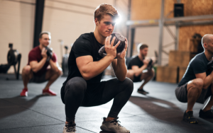
Why Squatting Triggers It
A squat isn't just one motion — it's a combination of deep hip flexion, internal rotation, and adduction happening simultaneously.
That specific triplanar pattern is so reliably provocative in FAI patients that clinicians use it as a diagnostic test (the FADIR test), which comes back positive in 88% of confirmed cases. In other words, squatting doesn't cause FAI, but it is almost perfectly designed to aggravate it.
The depth of the squat determines how much trouble you're in:
- Below 60° of flexion: Most FAI hips move without issue
- Between 60° and 90°: The femoral neck starts approaching the anterosuperior rim as the femur undergoes relative internal rotation
- Beyond 90°: Direct contact. A parallel squat demands roughly 110–120° of hip flexion; a full-depth squat pushes that to 140–160°
The research bears this out. FAI patients achieve 40% less pelvic range of motion during squats compared to people without the condition — 14.7° versus 24.2° in controls. They also show significantly reduced peak hip internal rotation at depth, averaging 9.4° against 15.2° in controls.
Motion analysis consistently shows these individuals unconsciously adopt protective strategies: shallower depth, reduced internal rotation, and greater external rotator activation to resist the impingement position.
Pelvic position adds another layer of complexity. The “butt wink” — posterior pelvic tilt at the bottom of the squat — actually increases anterior acetabular coverage, directly worsening impingement.
But the opposite extreme isn't safe either; excessive lumbar extension reduces hip joint space by 10–20%. FAI patients are essentially caught between two problematic positions.
Perhaps the most important thing to understand: this isn't a flexibility problem. These patients' bones physically collide before their muscles ever reach end-range. No amount of hip stretching changes that geometry.
How Clinicians Diagnose It
No single finding confirms FAI. A proper diagnosis requires all three elements together: symptoms, clinical signs, and imaging. Missing any one of them risks both over- and under-diagnosis.
Symptoms
The typical presentation is a gradual onset of deep groin pain that worsens with prolonged sitting, squatting, lunging, and rising from a chair. Athletes often notice their squat depth quietly decreasing over months before pain becomes the dominant complaint.
Clicking or catching in the joint points toward labral involvement. Strength testing commonly reveals deficits of up to 30% in the glutes, hip flexors, and adductors compared to the unaffected side — a detail that matters both for diagnosis and for building a rehab plan.
Clinical Testing
Clinicians rely on a cluster of provocation tests rather than any single maneuver:
- FADIR test (flexion to 90°, then adduction and internal rotation): Sensitivity of 80–96% makes it an excellent screening tool. A negative result effectively rules out FAI, though a positive result alone doesn't confirm it — specificity is poor at just 11–33%.
- FABER test (flexion, abduction, external rotation into a figure-4 position): Primarily useful for differentiating hip pathology from sacroiliac dysfunction based on where pain is reproduced.
- Log roll test (gently rolling the extended leg into internal and external rotation): More specific for intra-articular pathology because it minimizes soft-tissue involvement.
Imaging
Standard AP pelvis and lateral X-rays come first, identifying cam morphology, pincer morphology, and any joint space narrowing that might suggest early osteoarthritis.
MR arthrography is the gold standard when you need to visualize labral tears and cartilage damage in detail. CT scanning is reserved primarily for surgical planning, where three-dimensional bony detail becomes important.
A Critical Caveat on Differential Diagnosis
Groin pain in athletes has a long differential — adductor strains are actually the most common cause, identifiable by tenderness along the inner thigh and pain with resisted adduction.
Athletic pubalgia, iliopsoas tendinitis, labral tears without FAI morphology, and stress fractures all warrant consideration.
The complicating factor: roughly 86% of athletes with groin pain show radiographic FAI features, and multiple pathologies frequently coexist. Thorough evaluation isn't optional — it's what separates a targeted treatment plan from months of misdirected effort.
Conservative Management and Squat Modifications
Physical therapy is the starting point for FAI, and the outcomes data is encouraging. A meta-analysis of conservative interventions found moderate-to-large effect sizes for both pain reduction and functional improvement.
A study of 97 hips showed that 54.6% of patients achieved normal daily activity without ever needing surgery, and among younger athletes, 70% were successfully managed with therapy alone.
The Four Pillars of Rehab
Effective conservative management targets four areas: postural control, core stabilization, hip strength and motor control, and functional range of motion within pain-free limits. These aren't independent — they build on each other, and skipping ahead undermines the whole process.
One counterintuitive early insight: hip flexors that feel tight in FAI patients are usually compensating for poor core stability rather than being structurally shortened.
Stretching them without first addressing the underlying instability tends to worsen symptoms, not improve them. Core-first programming consistently outperforms strengthening-alone approaches for pain reduction.
Exercise Progression
Rehabilitation follows a logical sequence from non-weight-bearing toward full loading:
- Foundational: Glute bridges, clamshells, dead bugs, bird-dogs, and 90/90 breathing drills to establish core coordination
- Intermediate: Banded lateral walks, standing hip extension and abduction with resistance, single-leg Romanian deadlifts, step-ups, and lateral step-downs
- Advanced: Modified squats, hip hinge patterns, and sport-specific movements within tolerance
Exercises that combine deep hip flexion with internal rotation — deep squats, deep lunges, full-depth leg press, rowing machines — should be avoided or significantly modified throughout rehab.
Aggressive stretching into the impingement position is equally counterproductive regardless of how tight the hip feels. Bony anatomy doesn't respond to mobility work.
Squat Modifications
The goal isn't to stop squatting — it's to find a variation that loads the lower body without driving the femoral neck into the acetabular rim. Several adjustments make a meaningful difference:
- Variation: Front squats and goblet squats promote a more upright torso, reducing hip flexion demand at any given depth. Low-bar back squats require the most forward lean and are the most provocative variation for FAI.
- Stance width and foot angle: Wider stance with toes turned out roughly 30° suits people with retroverted hip sockets, promoting external rotation and reducing anterior impingement. Those with anteverted hips may do better with a narrower stance. A practical self-check: find the foot position that allows maximum hip flexion before the lower back rounds — that's likely your optimal width.
- Depth: Limiting to above parallel keeps hip flexion below the impingement threshold for most people. Box squats are particularly useful here since they provide a consistent physical reference that prevents gradual drift deeper over a set.
- Heel elevation: Plates or a slant board encourage a more upright torso and shift demand toward the quads while reducing hip flexion. The heel-elevated narrow-stance front squat and Spanish squats are two low-impingement alternatives worth exploring.
Three technique points deserve attention regardless of variation: maintain a 360° core brace rather than simply arching the lower back, keep knees tracking over the second toe to prevent valgus collapse, and address any ankle dorsiflexion restrictions — limited ankle mobility forces compensatory hip flexion that compounds impingement stress.
When Surgery Is the Right Call
Surgery isn't the first option — it's what comes after conservative management has been given a genuine chance and hasn't delivered. Clinical guidelines generally require 3–6 months of supervised physical therapy, activity modification, and an NSAID trial before arthroscopy is considered.
The other key threshold is a diagnostic intra-articular injection: if it provides temporary relief, it confirms the pain is coming from inside the joint, which helps justify the surgical route.
The Procedure
Hip arthroscopy has largely replaced open surgery for FAI. The procedure typically addresses three things:
- Femoroplasty: Reshaping the femoral head-neck junction to remove the cam bump
- Acetabuloplasty: Trimming excess rim bone to eliminate the pincer component
- Labral repair: Reattaching the labrum using suture anchors, which is strongly preferred over simply debriding the damaged tissue based on consistently superior outcomes data
Who Does Well and Who Doesn't
Patient selection is where surgical outcomes are really determined. The best candidates are younger patients with minimal cartilage damage and no radiographic signs of osteoarthritis.
Pre-existing high-grade cartilage damage is the single strongest predictor of poor outcomes — 82% of patients who eventually required total hip replacement had significant cartilage damage at the time of their original arthroscopy.
Timing also matters: surgery within six months of symptom onset consistently produces better results than delayed intervention.
The outcomes in well-selected patients are strong. A meta-analysis covering 1,981 hips found significant improvement across all patient-reported outcome measures, a pooled reoperation rate of just 5.5%, and ten-year joint preservation rates reaching 90.4%.
Return to Squatting After Surgery
Recovery follows a predictable sequence with clear squat-specific milestones:
- Weeks 4–6: Wall squats and mini-squats begin
- Weeks 8–12: Bodyweight squats with progressive depth
- Weeks 12–16: Loaded barbell squats, provided hip range of motion is adequate, strength deficits are below 30%, and movement is pain-free
- Months 4–6: Full-depth heavy squatting becomes reasonable
Mean return to sport across studies lands at 5–7 months, with 93% of patients returning to sport at some level and 82% returning to their pre-injury level of activity — figures that compare favorably to most other joint procedures.
Conclusion
FAI-related groin pain during squatting is a mechanical problem with a mechanical explanation — abnormal bone morphology eliminates the clearance your hip needs to flex deeply under load, and no amount of stretching or mobility work changes that geometry.
For most people, the combination of smart rehab and thoughtful squat modifications is enough to get back to training without surgery.
If it isn't, arthroscopy in the right candidate delivers reliable, durable results — but the single most actionable shift you can make right now is to stop chasing depth and start modifying your movement to match your anatomy.